
Clinical Cases Performed Under the Dental Microscope

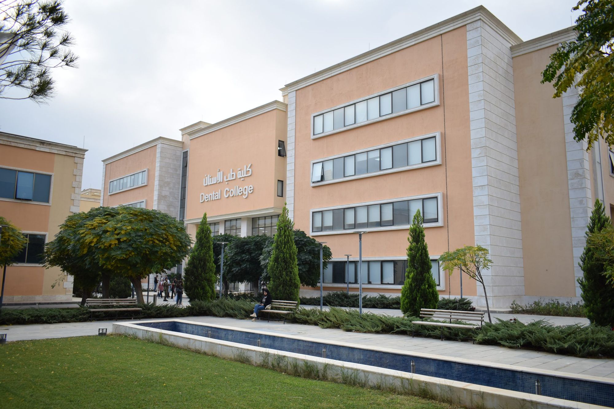
Clinical cases performed under microscopic magnification at the Faculty of Dentistry, Syrian Private University. The video of each case shows all its clinical stages.

01

Clinical case 1: Class III aesthetic restoration

Performed by Prof. Dr. Talal Al Nahlawi, Department of Operative Dentistry

A 25-year-old patient consulted the Faculty of Dentistry at the Syrian Private University complaining of temporary sensitivity to cold drinks that disappeared once the cause was removed.

Clinical and radiographic examination showed medium-depth caries on the distal surface of tooth No. 21 (FDI). The treatment plan was to remove the caries and place an aesthetic composite restoration under rubber dam isolation.

The carious lesion was prepared and restored under microscopic magnification; the attached video shows all the clinical stages.

Watch the case video
02

Clinical case 2: Endodontic treatment – rotary preparation and thermal obturation

Performed by Prof. Dr. Talaat Abo Hatab, Department of Operative Dentistry

A 23-year-old patient consulted the Faculty of Dentistry at the Syrian Private University complaining of severe, continuous spontaneous attacks of pain.

Clinical and radiographic examination showed deep, penetrating caries on the distal surface of tooth No. 15 (FDI). The treatment plan was to remove the inflamed pulp tissue through endodontic treatment under rubber dam isolation.

The pulp chamber was opened and the root canal was prepared, disinfected and filled with three-dimensional thermal obturation under microscopic magnification. The attached video shows all the clinical stages.

Watch the case video
03

Clinical case 3: Class I aesthetic restoration

Performed by Prof. Dr. Talal Al Nahlawi, Department of Operative Dentistry

A 22-year-old patient consulted the Faculty of Dentistry at the Syrian Private University complaining of temporary sensitivity to cold drinks that disappeared once the cause was removed.

Clinical and radiographic examination showed medium-depth caries on the occlusal surface of tooth No. 16 (FDI), the upper first molar, together with proximal caries. The treatment plan was to remove the caries and place an aesthetic composite restoration under rubber dam isolation, and to treat the proximal caries in a later, separate session.

The carious lesion was prepared and restored under microscopic magnification; the attached video shows all the clinical stages.

Watch the case video
04

Clinical case 4: Class II aesthetic restoration

Performed by Prof. Dr. Talal Al Nahlawi, Department of Operative Dentistry

An 18-year-old patient consulted the Faculty of Dentistry at the Syrian Private University complaining of temporary sensitivity to cold drinks and sweet foods that disappeared once the cause was removed, indicating reversible pulpitis.

Clinical and radiographic examination showed medium-depth caries on the proximal surface of tooth No. 16 (FDI), the upper first molar. The treatment plan was to remove the caries and place an aesthetic composite restoration under rubber dam isolation.

The carious lesion was prepared and restored under microscopic magnification; the attached video shows all the clinical stages.

Watch the case video
05

Clinical case 5: Endodontic treatment with rotary preparation and lateral condensation

Performed by fifth-year student Ahmad Abdul Ilah, supervised by Dr. Ahmad Al Abdullah, Department of Operative Dentistry

A 19-year-old patient consulted the Faculty of Dentistry at the Syrian Private University complaining of constant, continuous, radiating and throbbing pain, both spontaneous and provoked by cold and hot drinks.

Clinical and radiographic examination showed deep, penetrating caries on the distal surface of tooth No. 35 (FDI). The treatment plan was to remove the caries and perform endodontic treatment under rubber dam isolation.

The root canal was prepared with rotary instruments and filled by lateral condensation under microscopic magnification.

06

Clinical case 6: Endodontic treatment with rotary preparation and lateral condensation

Performed by fifth-year student Mohammad Osama Al Hamwi, supervised by Dr. Ahmad Al Abdullah, Department of Operative Dentistry

A 38-year-old patient consulted the Faculty of Dentistry at the Syrian Private University complaining of constant, continuous, radiating and throbbing pain, both spontaneous and provoked by cold and hot drinks.

Clinical and radiographic examination showed deep, penetrating caries on the distal surface of tooth No. 34 (FDI). The treatment plan was to remove the caries and perform endodontic treatment under rubber dam isolation.

The root canal was prepared with rotary instruments and filled by lateral condensation under microscopic magnification; the attached video shows all the clinical stages.

Watch the case video
07

Clinical case 7: Endodontic treatment with rotary preparation and lateral condensation

Performed by fifth-year student Tayseer Al Hamoud, supervised by Dr. Ahmad Al Abdullah, Department of Operative Dentistry

A 32-year-old patient consulted the Faculty of Dentistry at the Syrian Private University complaining of constant, continuous, radiating and throbbing pain, both spontaneous and provoked by cold and hot drinks.

Clinical and radiographic examination showed deep, penetrating caries on the distal surface of tooth No. 35 (FDI). The treatment plan was to remove the caries and perform endodontic treatment under rubber dam isolation.

The root canal was prepared with rotary instruments and filled by lateral condensation under microscopic magnification; the attached video shows all the clinical stages.

Watch the case video
08

Clinical case 8: Endodontic complication management – furcation perforation repair

Performed by Prof. Dr. Talaat Abo Hatab, Department of Operative Dentistry

A 27-year-old female patient consulted the Faculty of Dentistry at the Syrian Private University complaining of intermittent painful attacks and gingival swelling at one of her previously treated teeth.

Clinical and radiographic examination showed a perforation in the furcation area on the distal surface of tooth No. 14 (FDI). The treatment plan was to repair the perforation with MTA and complete the endodontic treatment under rubber dam isolation. All treatment procedures were carried out under micro-magnification using a dental microscope.

The attached video shows all the clinical stages.

Watch the case video